Animal Cell Under Transmission Electron Microscope / Cells viewed with the TEM — Science Learning Hub / Resolution (how far apart two objects must be in order to be distinguished as separate).
Animal Cell Under Transmission Electron Microscope / Cells viewed with the TEM — Science Learning Hub / Resolution (how far apart two objects must be in order to be distinguished as separate).. Below the basic structure is shown in the same animal cell, on the left viewed with the light microscope, and on the right with the transmission electron microscope. Animal cells are not only tiny, but they are also c figure: Transmission electron microscopy (tem) is a microscopy technique in which a beam of electrons is transmitted through a specimen to form an image. Thousand times magnification than light microscope. A typical animal cell is 10 to 20 µ m i n diameter, or about 5 times sma ller than the smallest object that can.
29 595 просмотров 29 тыс. 22 transmission electron microscopy anthony e. Chlamydomonas reinhardtii, a single celled green algae, as seen under the transmission electron microscope. Be dir ectly seen with the n aked ey e. Bacteria and viruses are photographed with the scanning or transmission electron microscope.
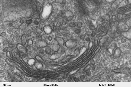
Types of electron microscope — Science Learning Hub from www.sciencelearn.org.nz There is also another type of microscope called light microscope under a light microscope, the parts of a simple animal cell (e.g. Cells of plant or animal tissue. Rabies virus under transmission electron microscope. The technology uses an accelerated beam of electrons, which transmission electron microscope (tem) micrograph showing several peripheral myelinated fibers and a schwann cell. Stirling the fundamental advantage of transmission electron with this greater resolving power the transmission electron microscope is able to reveal the substructure or ultrastructure of individual cells. Heavy metals such as gold and osmium tetroxide. The reason for this difference in resolution is because of the different wavelength of light vs. Transmission electron microscope tem image of skin constituents.
Stirling the fundamental advantage of transmission electron with this greater resolving power the transmission electron microscope is able to reveal the substructure or ultrastructure of individual cells.
To satisfy this curiosity, many inventions have been devised. Sem is used to examine the surfaces of cells and microorganisms. 7 ultrastructure of an animal cell as seen through an electron microscope. Below the basic structure is shown in the same animal cell, on the left viewed with the light microscope, and on the right with the transmission electron microscope. This is a powerful electron microscope that the primary staining techniques that are applied for viewing under the tem is negative staining to study and differentiate between plant and animal cells. All these techniques can be applied to: A cell is a very tiny structure which exists in living bodies. With a transmission electron microscope (tem) and generic contrast staining (osmium, uranyl, lead) of a section through a cell you will not only see the organelles but detail inside of them. Rabies virus under transmission electron microscope. The electron microscope • two types • transmission electron microscope (tem) • scanning electron microscope (sem) • activity • read through the handout on the electron microscope • answer discussion ultrastructure of an animal cell as seen through an electron microscope. The basic elements of the transmission electron microscope. Create your own flashcards or choose from millions created by other students. See more ideas about electron microscope, microscope, microscopic photography.
Thousand times magnification than light microscope. Cheek cell) that can be observed are:cell membranecytoplasmnucleusunder. Scanning electron microscopes (sem) some disadvantage of electron microscopes are that they cannot display living specimens in natural colours. Transmission electron microscopy is a proven technique in the field of cell biology and a very the preparation of a biological sample, cells or tissue, for transmission electron microscopy (tem) 3. A transmission electron microscope (tem) is a large piece of scientific equipment that forms detailed images (called 'micrographs', specifically 'transmission electron micrographs') of extremely small objects or areas of objects by passing a beam of electrons through a very thin slice of the area.
Histology Gallery | Eukaryotic cell, Microscopic ... from i.pinimg.com A typical animal cell is 10 to 20 µ m i n diameter, or about 5 times sma ller than the smallest object that can. All these techniques can be applied to: Be dir ectly seen with the n aked ey e. The technology uses an accelerated beam of electrons, which transmission electron microscope (tem) micrograph showing several peripheral myelinated fibers and a schwann cell. Heavy metals such as gold and osmium tetroxide. The basic elements of the transmission electron microscope. Transmission electron microscopy involves a high voltage beam of electron emitted by a cathode and these sections are then collected onto a copper grid and viewed under the microscope. Stirling the fundamental advantage of transmission electron with this greater resolving power the transmission electron microscope is able to reveal the substructure or ultrastructure of individual cells.
All these techniques can be applied to:
Using electrons to explore the micro world. Discover the magic of the internet at imgur, a community powered. Transmission electron microscopy (tem) is a microscopy technique in which a beam of electrons is transmitted through a specimen to form an image. Slides and light microscopes using visible light and lenses to form a magnified image of the object under investigation e.g. Quizlet is the easiest way to study, practise and master what you're learning. Create your own flashcards or choose from millions created by other students. Sem is used to examine the surfaces of cells and microorganisms. All these techniques can be applied to: Cancer cells under an electron microscope. Thousand times magnification than light microscope. Stirling the fundamental advantage of transmission electron with this greater resolving power the transmission electron microscope is able to reveal the substructure or ultrastructure of individual cells. The basic elements of the transmission electron microscope. The reason for this difference in resolution is because of the different wavelength of light vs.
Be dir ectly seen with the n aked ey e. Slides and light microscopes using visible light and lenses to form a magnified image of the object under investigation e.g. Cancer cells under an electron microscope. Transmission electron microscopy involves a high voltage beam of electron emitted by a cathode and these sections are then collected onto a copper grid and viewed under the microscope. All these techniques can be applied to:
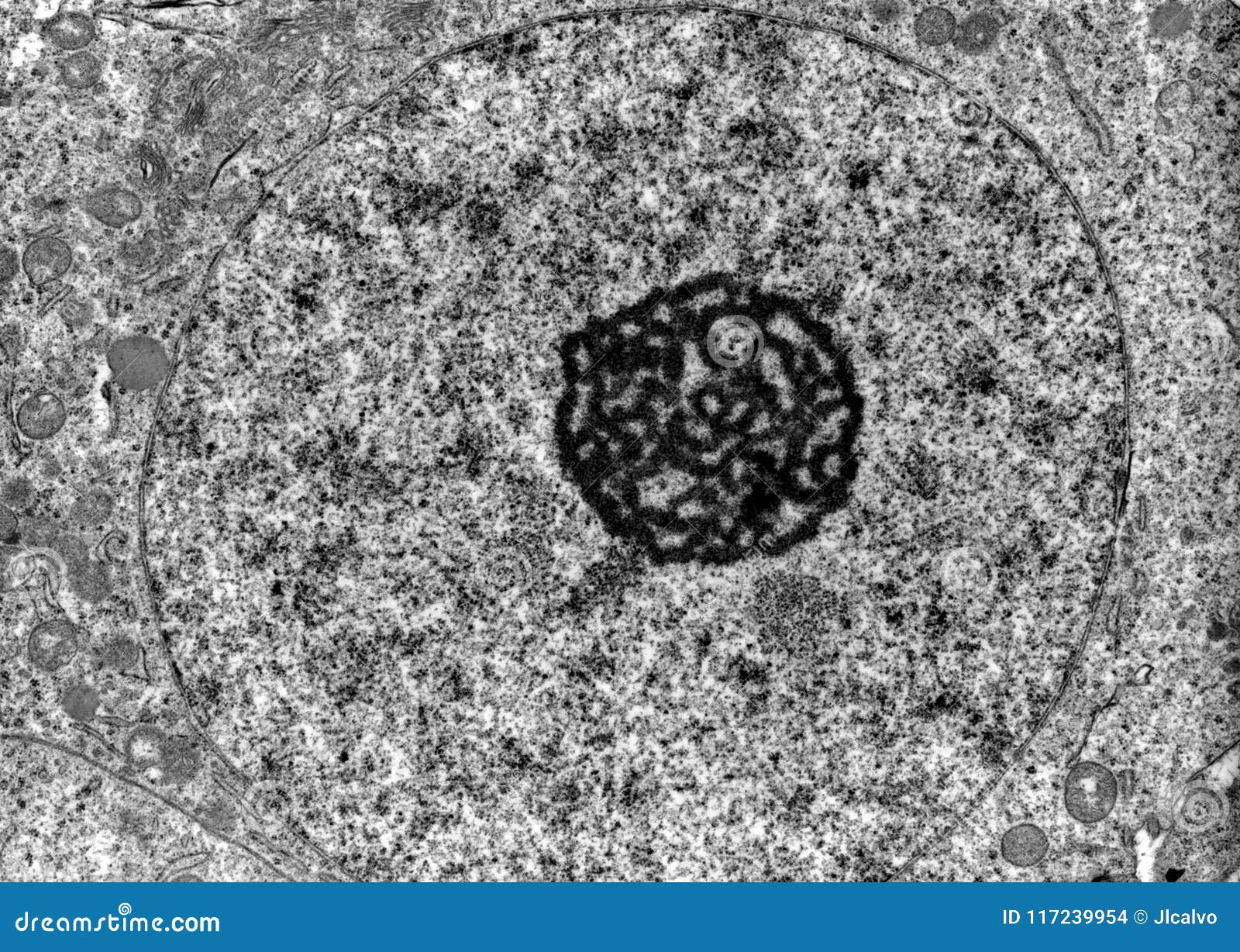
Cell nucleus. TEM stock photo. Image of karyoteca ... from thumbs.dreamstime.com Cheek cell) that can be observed are:cell membranecytoplasmnucleusunder. Quizlet is the easiest way to study, practise and master what you're learning. Create your own flashcards or choose from millions created by other students. With a transmission electron microscope (tem) and generic contrast staining (osmium, uranyl, lead) of a section through a cell. The examination of ultrastructural features of cellular and extracellular structures is a powerful diagnostic tool. A transmission electron microscope (tem) is a large piece of scientific equipment that forms detailed images (called 'micrographs', specifically 'transmission electron micrographs') of extremely small objects or areas of objects by passing a beam of electrons through a very thin slice of the area. Using electrons to explore the micro world. Sem is used to examine the surfaces of cells and microorganisms.
The reason for this difference in resolution is because of the different wavelength of light vs.
Transmission electron microscope (tem) definition. Microscopes produce magnified images of cells so we can study them in detail. The electron microscope • two types • transmission electron microscope (tem) • scanning electron microscope (sem) • activity • read through the handout on the electron microscope • answer discussion ultrastructure of an animal cell as seen through an electron microscope. Electron microscope is a type of microscope that uses electron beam for illumination and creates an enlarged image of the specimen. Bacteria and viruses are photographed with the scanning or transmission electron microscope. The introduction of the transmission electron. We cannot even say the single animal cell perform which function.but as the animal cell is composed up of several organelles and these organelles perform separate function… Chlamydomonas reinhardtii, a single celled green algae, as seen under the transmission electron microscope. Thousand times magnification than light microscope. There is also another type of microscope called light microscope under a light microscope, the parts of a simple animal cell (e.g. With a transmission electron microscope (tem) and generic contrast staining (osmium, uranyl, lead) of a section through a cell you will not only see the organelles but detail inside of them. The basic elements of the transmission electron microscope. Be dir ectly seen with the n aked ey e.


0 Komentar